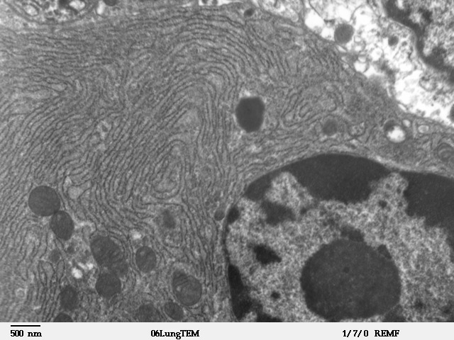
فَیِل:Clara cell lung - TEM.jpg
Clara_cell_lung_-_TEM.jpg (640 × 480 پِکسَل، فَیِل ناپ: 98 کِلوبایِٹ، MIME قسٕم:image/jpeg)
فَیِل تَوٲریٖخ
فَیِل وُچھنہٕ باپتھ کٔریو کلک تأریخ/وقت پؠٹھ تاکہِ یہ گژھِ تمہ وقتہٕ ظٲہر
| تٲریٖخ/وَقت | تھمب نیل | پہلوٗو | صٲرِف | کَتھ | |
|---|---|---|---|---|---|
| موجودٕ | 20:39, 4 اَکتوٗبَر 2006 |  | 640 × 480 (98 کِلوبایِٹ) | Patho | {{Information |Description=Transmission electron microscope image of a thin section cut through an area of mammalian lung tissue. This image of a Clara cell shows a nucleus and cytoplasmic organelles, such as rough endoplasmic reticulum and mitochondria. |
فَیِلٕ ہُند اِستِعمال
یہِ صَفہٕ چھُ اَتھ فَیِلہِ اِستِمال کَران:
فَیِلہٕ ہُنٛد عالمِی اِستِمال
دِنہٕ آمٕتیٚو باقٕی وِکیٖیَن منٛز چھےٚ یہِ بٕہی استعمال سپدان:
- ar.wikipedia.org پؠٹھ استعمال
- ckb.wikipedia.org پؠٹھ استعمال
- cs.wikipedia.org پؠٹھ استعمال
- de.wikipedia.org پؠٹھ استعمال
- de.wikibooks.org پؠٹھ استعمال
- el.wikipedia.org پؠٹھ استعمال
- en.wikipedia.org پؠٹھ استعمال
- eo.wikipedia.org پؠٹھ استعمال
- fa.wikipedia.org پؠٹھ استعمال
- gl.wikipedia.org پؠٹھ استعمال
- gv.wikipedia.org پؠٹھ استعمال
- ht.wikipedia.org پؠٹھ استعمال
- hu.wikipedia.org پؠٹھ استعمال
- hy.wikipedia.org پؠٹھ استعمال
- it.wikibooks.org پؠٹھ استعمال
- jv.wikipedia.org پؠٹھ استعمال
- kn.wikipedia.org پؠٹھ استعمال
- mk.wikipedia.org پؠٹھ استعمال
- ml.wikipedia.org پؠٹھ استعمال
- mn.wikipedia.org پؠٹھ استعمال
- ms.wikipedia.org پؠٹھ استعمال
- pl.wikibooks.org پؠٹھ استعمال
- ru.wikibooks.org پؠٹھ استعمال
- sh.wikipedia.org پؠٹھ استعمال
- simple.wikipedia.org پؠٹھ استعمال
- si.wikipedia.org پؠٹھ استعمال
- sl.wikipedia.org پؠٹھ استعمال
- sr.wikipedia.org پؠٹھ استعمال
- ta.wikipedia.org پؠٹھ استعمال
- th.wikipedia.org پؠٹھ استعمال
- tl.wikipedia.org پؠٹھ استعمال
- uk.wikipedia.org پؠٹھ استعمال
- ur.wikipedia.org پؠٹھ استعمال
